Short Courses2
Short Course 2: Data Science and AI Models in Precision Cancer Research
May 15, 2025, 1:00pm-5:00pm PDT
Stanford Campus, Always M121Q
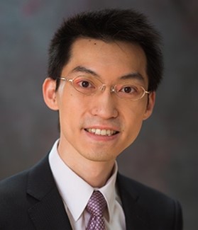
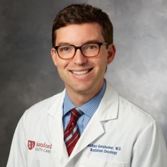
Instructor: Lei Xing, Stanford University; Kun-Hsing Yu, Harvard University; Ruogu Fang, University of Florida, and Michael Gensheimer, Stanford University.
 Lei Xing, PhD, the Jacob Haimson & Sarah S. Donaldson Professor of Medical Physics and Electrical Engineering (by courtesy) at Stanford University. He is also the Director of the Medical Physics Division of Radiation Oncology Department. His research is focused on AI in medicine, medical imaging, and biomedical physics. He has made unique and significant contributions to each of the above areas. Dr. Xing is an author on more than 450 peer reviewed publications, a co-inventor on many issued and pending patents, and a co- investigator or PI on numerous NIH, NSF, DOD, RSNA, ACS and many corporate grants. He is a fellow of AAPM (American Association of Physicists in Medicine), ASTRO (American Society for Radiation Oncology), and AIMBE (American Institute for Medical and Biological Engineering). He has received numerous awards, such as Google Faculty Scholar Award and E. H. Quimby Lifetime Achievement Awards from AAPM.
 "Kun-Hsing "Kun" Yu, MD, PhD, is an Assistant Professor of Biomedical Informatics, Harvard Medical School. He developed the first fully automated artificial intelligence (AI) algorithm to extract thousands of features from whole-slide histopathology images, discovered the molecular mechanisms underpinning the microscopic phenotypes of tumor cells, and successfully identified previously unknown cellular morphologies associated with patient prognosis. His lab integrates cancer patients' multi-omics (genomics, epigenomics, transcriptomics, and proteomics) profiles with quantitative histopathology patterns to predict their clinical phenotypes. More than 30 research laboratories worldwide have independently validated the AI methods developed in the Yu Lab. Dr. Yu's work in pathology AI has received several accolades, including the American Medical Informatics Association New Investigator Award, the Department of Defense Career Development Award, the Google Research Scholar Award, and the National Institutes of Health (NIH) Maximizing Investigators' Research Award. He is a Fellow of the American Medical Informatics Association.
 Ruogu Fang, PhD, tenured Associate Professor and Pruitt Family Endowed Faculty Fellow in the J. Crayton Pruitt Family Department of Biomedical Engineering at the University of Florida. Her research encompasses two principal themes: AI-empowered precision brain health and brain/bio-inspired AI. Her work involves addressing compelling questions, such as using machine learning techniques to quantify brain dynamics, facilitating early Alzheimer's disease diagnosis through novel imagery, predicting personalized treatment outcomes, designing precision interventions, and leveraging principles from neuroscience to develop the next-generation of AI. Fang's current research is also rooted in the confluence of AI and multimodal medical image analysis. She is the PI of an NIH NIA RF1 (R01-equivalent), an NSF Research Initiation Initiative (CRII) Award, an NSF CISE IIS Award, a Ralph Lowe Junior Faculty Enhancement Award from Oak Ridge Associated Universities (ORAU). She has also received numerous recognitions. She was selected as the Rising Stars (Engineering) by the Academy of Science, Engineering, and Medicine of Florida (ASEMFL), the inaugural recipient of the Robin Sidhu Memorial Young Scientist Award from the Society of Brain Mapping and Therapeutics, an Best Paper Award from the IEEE International Conference on Image Processing , the University of Florida AI Course Award, an UF Herbert Wertheim College of Engineering Faculty Award for Excellence in Innovation, an UF BME Faculty Research Excellence Award and Faculty Teaching Excellence Award, among others. Fang's research has been featured by Forbes Magazine, The Washington Post, ABC, RSNA, and published in Lancet Digital Health, JAMA, PNAS and npj Digital Medicine. She is an Associate Editor of the Journal Medical Image Analysis, a Topic Editor of Frontiers in Human Neuroscience, and a Guest Editor of CMIG. She is a reviewer for The Lancet, Nature Machine Intelligence, Science Advances, etc. Her research has been supported by NSF, NIH, Oak Ridge Laboratory, DHS, DoD, NVIDIA, and the University of Florida. She is President of Women in MICCAI (WiM) and Associate Editor of the Medical Image Analysis journal. At the heart of her work is the Smart Medical Informatics Learning and Evaluation (SMILE) lab, where she is tirelessly dedicated to creating groundbreaking brain and neuroscience-inspired medical AI and deep learning models. The primary objective of these models is to comprehend, diagnose, and treat brain disorders, all while navigating the complexities of extensive and intricate datasets.
 Michael Gensheimer, MD, Clinical Associate Professor of Radiation Oncology, Stanford University. He is a radiation oncologist whose clinical practice focuses on treatment of head and neck cancer. Much of his research involves analysis of large-scale electronic medical record datasets to predict patient outcomes and pick the most effective treatments. His predictive models have been deployed at Stanford and tested in randomized studies. His open source nnet-survival software that allows use of neural networks for survival modeling has been used by researchers internationally.
Abstract
Data science and AI models have gained popularity in medical research. This short course provides an introduction and demonstration of these advanced models in cancer research. We will start with an introduction of foundation models and how they will be related to individual medicine. An imaging foundation models for neurodegenerative disease will be presented to illustrate the development and application of foundation models in medicine. We will than explain the use of large language models in precision oncology and oncology practice through examples. Finally, we will present the cutting-edge research in a multimodality AI for next generation of biomedicine provides power tools but also provides a lot of opportunities in research and applications in clinical oncology.